- Chris Comans
- 0 Comments
The skin around the eyes is among the most delicate on the body. It is thin, highly exposed to ultraviolet (UV) radiation, and often neglected when applying sunscreen. Decades of sunbathing with eyes closed provided no real protection — ultraviolet rays penetrated the eyelids, causing long-term cellular damage. This explains why basal cell carcinoma (BCC), squamous cell carcinoma (SCC), and ocular melanoma can appear in this region.
At Skin ChX Subiaco, Christine Comans, Dermal Clinician and Skin Cancer Screening Practitioner, provides detailed skin cancer screening with dermatoscopic assessment, including the periocular region.
However, because intraocular structures cannot be evaluated during a skin check, it remains vital that patients also attend regular eye examinations with an optometrist.
Optometrists use specialised equipment to examine inside the eye for early signs of melanoma or other eye disease, complementing the work done at skin checks.
Ocular Melanoma
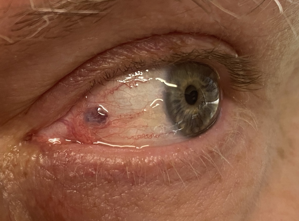
Ocular melanoma arises from melanocytes within the eye but may also be visible on the conjunctiva or eyelid margin.
Dermatoscopic features include:
- Structureless pigmentation in brown, grey, or black.
- Blue pigmentation caused by the Tyndall effect, where light scatter in the dermis makes brown pigment appear blue.
- Polychromia, with multiple tones (light brown, dark brown, grey, black).
- Absence of a pigment network, unlike benign conjunctival naevi.
While a dermatoscopic skin check can detect melanomas affecting the eyelid margin, only an optometrist or ophthalmic specialist can assess the internal eye. This underlines the importance of combining eye exams with skin cancer screening.
Basal Cell Carcinoma (BCC) Around the Eye
Basal cell carcinoma is the most common eyelid cancer. Dermatoscopy helps to identify its features early, even when clinical signs are subtle.
Common dermatoscopic findings are:
- Arborising telangiectasia: Branching blood vessels of varying calibre.
- Blue-grey ovoid nests: Well-defined pigmented structures representing tumour cell aggregates.
- Shiny white areas: Seen under polarised light, indicating dermal fibrosis.
- Structureless ulceration or erosion, sometimes with haemorrhage.
Periocular BCC may be mistaken for a stye or chalazion. Dermatoscopy provides clarity by revealing features not found in inflammatory conditions.
Squamous Cell Carcinoma (SCC) of the Eyelid
SCC carries greater metastatic risk than BCC and can grow rapidly if undetected.
Dermatoscopic features include:
- White-yellow structureless areas, consistent with keratin.
- Surface scaling and crusting, often concentrated centrally.
- Polymorphous vascular patterns, including dotted, linear irregular, hairpin, and glomerular vessels.
- Whitish perivascular halos around some vessels.
- Pink structureless backgrounds, producing an irregular, heterogeneous appearance.
Eyelid SCC is often overlooked because patients believe they have conjunctivitis, blepharitis, or minor irritation. This can delay diagnosis. Past sunbathing without adequate eye protection remains a significant risk factor.
Why Eye Exams and Skin Checks Go Together
- Optometrists assess the internal structures of the eye, including the retina and choroid, where melanomas may arise beyond the reach of dermatoscopy.
- Skin checks at Skin ChX Subiaco focus on the external structures — the eyelids, periocular skin, and surrounding face — detecting BCC, SCC, and conjunctival melanomas.
- When both forms of screening are performed regularly, the chance of missing a significant lesion is reduced.
Key Messages
- Ocular melanoma: Dermatoscopy shows structureless pigmentation, polychromia, and Tyndall blue areas, but internal melanomas require optometric assessment.
- Basal cell carcinoma: Arborising vessels, blue-grey nests, shiny white areas, and ulceration are typical dermatoscopic findings.
- Squamous cell carcinoma: White-yellow keratin, polymorphous vascularity, and pink backgrounds are common features.
- Delayed recognition is common: Many patients mistake eyelid cancers for irritation or infection.
- Dual approach: Regular eye exams with an optometrist plus skin checks at Skin ChX Subiaco provide comprehensive protection.
Conclusion
Periocular cancers are diagnostically challenging due to subtle clinical signs and overlapping features with benign conditions. Dermatoscopy improves accuracy by revealing vascular and structural features typical of malignancy. However, dermatoscopy cannot evaluate the inside of the eye — for this, optometric examinations are essential.
At Skin ChX Subiaco, Christine Comans integrates dermatoscopic skin checks into routine screening, ensuring that eyelid and periocular skin cancers are not overlooked. By pairing these skin checks with regular eye examinations, patients can feel confident that both the external and internal structures of the eye are being safeguarded.